
The Complete Clinical Guide to the Human Digestive System: Architecture, Physiology, and Systemic Impact
A comprehensive clinical breakdown of the human digestive system. Discover how the gastrointestinal tract, accessory organs, and enteric nervous system govern human health.
disclosure This article contains affiliate links. Purchases may earn us a commission at no extra cost to you. We only recommend products we've reviewed.
The Complete Clinical Guide to the Human Digestive System: Architecture, Physiology, and Systemic Impact
Have you ever considered what actually happens to the food you eat after the simple act of swallowing? Most people view human digestion as a passive, mechanical process—a basic biological tube designed to dissolve meals, extract basic calories, and discard remaining waste. However, modern neuro-gastroenterology and clinical physiology reveal a vastly different reality. The human digestive system is an extraordinarily sophisticated, highly autonomous metabolic and neural network that governs far more than hunger and bowel movements.
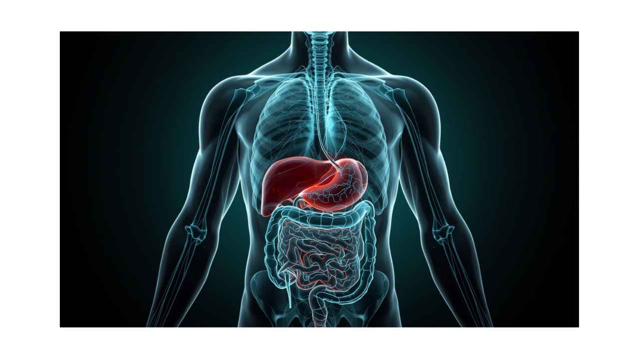
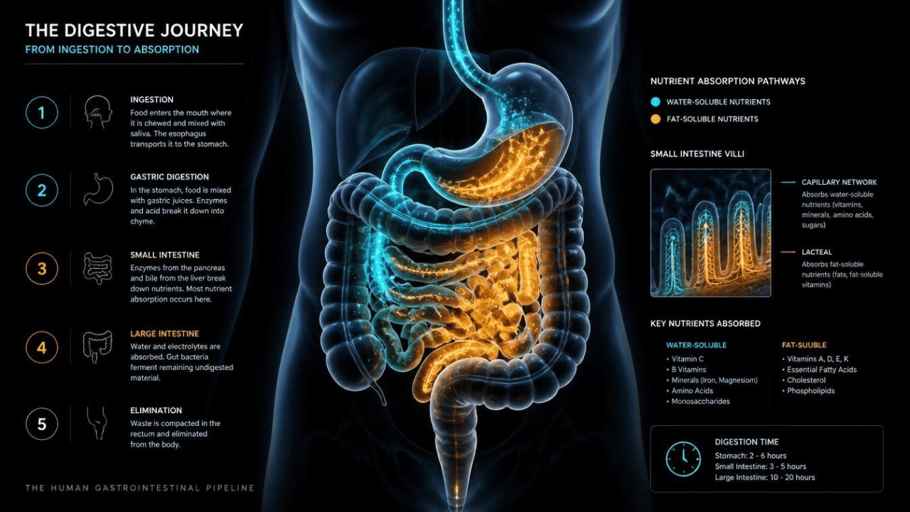 Conceptual 3D rendering of the human digestive pipeline, showing the continuous physiological route from the esophagus to the colon.
Stretching over twenty-five feet from the oral cavity to the rectum, this intricate internal pipeline interacts directly with every major system in your body. It processes tons of organic material over a lifetime, acts as your largest immune interface, and hosts trillions of living microorganisms that actively regulate your metabolism, hormone balance, and systemic inflammation. Furthermore, embedded deep within its cellular walls lies a dedicated neural system containing more than 500 million neurons, communicating continuously with your central nervous system.
Understanding the true architecture and physiology of the human digestive system is not merely an academic exercise. It is the absolute cornerstone of resolving chronic fatigue, persistent brain fog, metabolic dysfunction, and systemic autoimmune responses. In this comprehensive guide, we will explore every major stage of the digestive continuum, examining how each organ operates, how accessory tissues support chemical breakdown, and how the entire system functions as a unified biological engine.
1. The Upper Gastrointestinal Tract: Mechanical Breakdown and Acidification
The digestive journey begins long before food ever reaches your stomach. It starts in the oral cavity, where both mechanical and chemical digestion initiate simultaneously. Teeth perform mechanical mastication, physically breaking down complex food structures into smaller particles to dramatically increase their surface area. Concurrently, three major pairs of salivary glands—the parotid, submandibular, and sublingual glands—secrete a complex fluid rich in water, electrolytes, mucus, and crucial digestive enzymes.
Among these enzymes, salivary amylase initiates the chemical cleavage of complex starches into simpler sugars, while lingual lipase begins the preliminary breakdown of dietary lipids. The tongue then shapes this lubricated mixture into a soft, cohesive mass known as a bolus, pushing it toward the pharynx. The act of swallowing triggers a highly coordinated reflex: the soft palate rises to prevent food from entering the nasal cavity, while the epiglottis folds down over the larynx to protect the airway, directing the bolus safely into the esophagus.
The esophagus is a muscular tube approximately twenty-five centimeters long that acts as a dedicated transport corridor. Rather than relying on gravity, the esophageal wall utilizes organized, wave-like muscular contractions termed peristalsis. This process combines an outer layer of longitudinal smooth muscle and an inner layer of circular smooth muscle to push the bolus downward toward the stomach. At the terminal end of the esophagus lies the lower esophageal sphincter, a specialized ring of smooth muscle that acts as a tight unidirectional valve. This valve relaxes to allow the bolus into the stomach and immediately contracts to prevent highly acidic gastric juices from rising into the delicate esophageal mucosa.
When the bolus enters the stomach, it encounters a harsh, highly specialized biochemical environment. The gastric mucosa contains millions of deep invaginations known as gastric pits, which house specialized secretory cells. Parietal cells secrete concentrated hydrochloric acid, dropping the stomach's internal pH to an extraordinarily acidic level between 1.5 and 2.5. This intense acidity serves multiple crucial functions: it neutralizes ingested pathogens, denatures complex dietary proteins, and converts inactive pepsinogen—secreted by neighboring chief cells—into its active protein-cleaving form, pepsin.
Simultaneously, the stomach wall utilizes three distinct muscular layers—circular, longitudinal, and oblique—to vigorously churn and mix the ingested food with gastric secretions. Over several hours, the solid bolus is transformed into a uniform, highly acidic semiliquid substance known as chyme. Small, controlled pulses of chyme are then gradually released through the pyloric sphincter into the initial segment of the small intestine, ensuring that downstream organs are not overwhelmed by excessive volume or acidity.
2. The Accessory Powerhouses: Enzymatic Mastery and Biliary Secretions
While the gastrointestinal tract forms the physical tube through which food travels, chemical digestion could not be completed without the vital contributions of three accessory organs: the pancreas, the liver, and the gallbladder. These organs sit adjacent to the digestive tract and deliver powerful chemical agents directly into the duodeno-jejunal junction in response to hormonal triggers.
The pancreas is an essential dual-function gland situated deep in the upper abdomen. Its exocrine tissue produces over a liter of concentrated pancreatic juice each day, which is rich in bicarbonate ions and a wide spectrum of digestive enzymes. When acidic chyme enters the duodenum, it triggers the release of secretin and cholecystokinin, hormones that signal the pancreas to discharge its secretions. The high concentration of bicarbonate neutralizes the incoming hydrochloric acid, raising the luminal pH to an optimal level for enzymatic activity while protecting the intestinal lining from acid erosion.
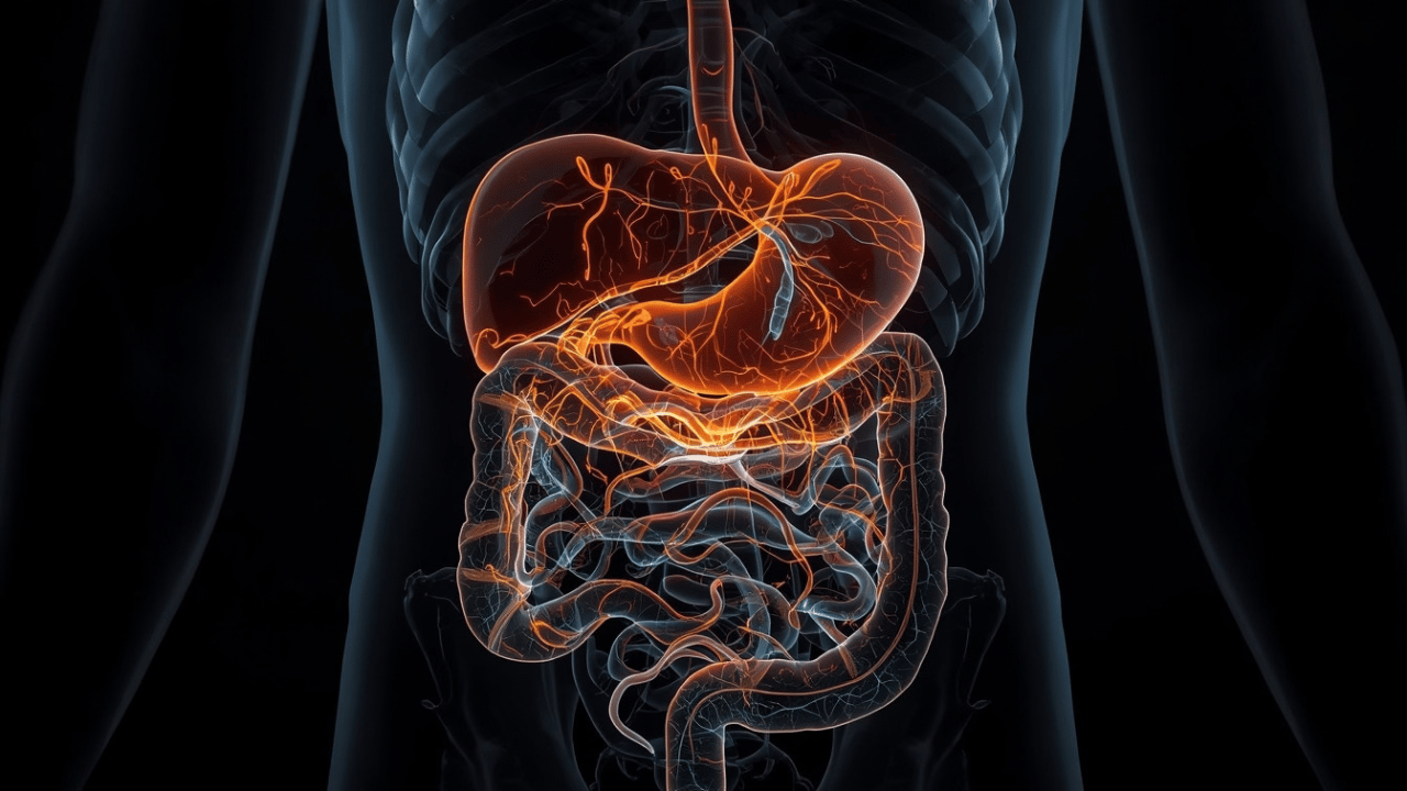
Clinical 3D medical visualization of the upper digestive hub, showing gastric acid secretion alongside the liver, gallbladder, and pancreas delivering enzymes into the duodenum.
The digestive enzymes produced by the pancreas represent the true heavy machinery of chemical breakdown. Pancreatic amylase continues the hydrolysis of complex carbohydrates into disaccharides. Pancreatic lipase, aided by specific co-factors, breaks down dietary triglycerides into free fatty acids and monoglycerides. Meanwhile, powerful proteolytic zymogens, including trypsinogen and chymotrypsinogen, are secreted in inactive forms and activated only within the duodenal lumen to safely cleave complex proteins into short peptide chains without digesting pancreatic tissue itself.
Located in the upper right quadrant of the abdomen, the liver is the largest internal organ in the human body and serves as a master metabolic processing laboratory. From a digestive standpoint, its primary role is the continuous production of bile—a complex fluid composed of bile salts, phospholipids, cholesterol, bilirubin, and electrolytes. Bile does not contain digestive enzymes; instead, its bile salts act as natural biological detergents. They emulsify large, insoluble fat globules into microscopic droplets, vastly increasing the surface area accessible to water-soluble pancreatic lipase.
Because the liver produces bile continuously, the body requires a storage facility to concentrate and release this fluid on demand. That role is fulfilled by the gallbladder, a small pear-shaped sac tucked beneath the liver. The gallbladder absorbs water and inorganic salts from hepatic bile, concentrating it up to tenfold. When fat-rich chyme enters the duodenum, the hormone cholecystokinin causes the gallbladder to contract vigorously, releasing concentrated bile through the common bile duct directly into the duodenal lumen to ensure efficient fat digestion and fat-soluble vitamin absorption.
3. The Small Intestine: The Primary Engine of Nutrient Absorption
Stretching approximately twenty feet in length, the small intestine represents the undisputed masterpiece of human digestive physiology. It is divided into three distinct anatomical regions, each optimized for specific biochemical tasks: the duodenum, the jejunum, and the ileum. While preliminary digestion occurs in the stomach, the vast majority of chemical breakdown and almost all nutrient absorption take place within this remarkable organ.
The first section, the duodenum, measures approximately twenty-five centimeters in length and acts as a central chemical processing hub. Here, acidic chyme from the stomach is thoroughly mixed with alkaline pancreatic juice and concentrated bile from the gallbladder. Specialized Brunner's glands located in the duodenal submucosa secrete an abundance of alkaline mucus to further neutralize gastric acid. Once the chyme is buffered and emulsified, it moves smoothly into the jejunum.
The jejunum spans roughly eight feet and serves as the primary site of nutrient absorption. To absorb huge quantities of amino acids, simple sugars, fatty acids, vitamins, and minerals, the internal surface area of the jejunum is amplified to an extraordinary degree through three hierarchical levels of anatomical engineering:
First, the luminal wall features permanent circular mucosal folds known as plicae circulares. Second, covering these folds are millions of tiny, finger-like projections called villi, each containing a dense network of blood capillaries and a central lymphatic vessel known as a lacteal. Third, the outer surface of each individual epithelial cell covering a villus is covered in thousands of microscopic extensions called microvilli, collectively forming the brush border. Combined, these structural adaptations expand the total absorptive surface area of the small intestine to roughly two hundred square meters—equivalent to the surface area of a tennis court.
At the brush border, specialized enzymes embedded directly within the cellular membranes complete the final stages of digestion. Disaccharidases such as lactase, sucrase, and maltase break down complex sugars into single glucose, galactose, and fructose molecules. Peptidases degrade short peptide chains into individual amino acids and dipeptides. Water-soluble nutrients are actively transported across the enterocytes and directly into the capillary blood stream, traveling straight to the liver via the hepatic portal vein. Conversely, re-esterified dietary fats and fat-soluble vitamins are packaged into lipid transport particles called chylomicrons and absorbed into the lymphatic lacteals, bypassing immediate liver filtration.
The final segment of the small intestine is the ileum, which stretches roughly twelve feet before joining the large intestine. While the jejunum handles the bulk of macronutrient absorption, the ileum is specialized for highly specific physiological tasks. It absorbs key micronutrients that require complex transport mechanisms, such as Vitamin B12 bound to intrinsic factor. Furthermore, the terminal ileum actively reabsorbs approximately ninety-five percent of the bile salts released into the digestive tract, returning them to the liver via the enterohepatic circulation to prevent biological waste and ensure continuous biliary capacity.
4. The Large Intestine and Microscopic Barrier Mechanics
Having passed through the entire length of the small intestine, the remaining unabsorbed material passes through the ileocecal valve and enters the large intestine. Measuring approximately five feet in length, the large intestine is shorter but significantly wider than the small intestine. It consists of the ceco-appendix, the colon (comprising the ascending, transverse, descending, and sigmoid regions), the rectum, and the anal canal.
Unlike the small intestine, the large intestine does not feature villi or produce digestive enzymes. Its primary mechanical and physiological responsibilities center on the absorption of water, the regulation of electrolyte balance, the fermentation of indigestible dietary fibers, and the consolidation of solid waste for elimination. As liquid chyme enters the cecum, slow haustral churning movements expose the material to the mucosal wall, allowing specialized epithelial cells to actively reabsorb sodium and water, gradually transforming liquid residues into formed stool.
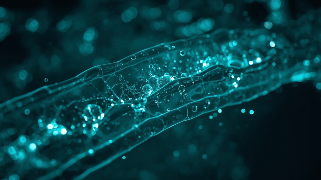
High-magnification clinical 3D rendering of the mucosal barrier lining, demonstrating tight junctions and nutrient absorption micro-channels in the intestinal epithelium.
However, the defining feature of the large intestine is not its mechanical tissue, but the staggering ecological ecosystem it houses: the gut microbiome. The human colon is home to over one hundred trillion living microorganisms, comprising hundreds of distinct bacterial species, archaea, and fungi. This complex microbial community functions as a highly active metabolic organ inside your body, processing complex dietary fibers and resistant starches that human digestive enzymes are incapable of breaking down.
Through anaerobic fermentation, beneficial gut bacteria transform these complex carbohydrates into short-chain fatty acids, primarily acetate, propionate, and butyrate. Butyrate serves as the primary energy source for the epithelial cells lining the colon, driving cellular repair, suppressing localized tissue inflammation, and reinforcing the integrity of tight junction proteins that seal adjacent intestinal cells together. Furthermore, the gut microbiota synthesizes essential micronutrients, including Vitamin K and several B-complex vitamins, while outcompeting potential pathogenic microbes for nutrients and biological binding sites.
The mechanical mucosal wall of the large intestine acts as a vital physiological barrier. It separates the trillions of microbes and toxic waste products residing inside the intestinal cavity from the sterile bloodstream and immune tissue sitting just fractions of a millimeter away. When chronic tissue inflammation, poor dietary factors, or unmanaged stress degrade this delicate mucosal barrier, tight junction proteins break down. This increase in intestinal permeability—clinically referred to as mucosal barrier breakdown—allows bacterial endotoxins like lipopolysaccharides to leak directly into systemic circulation, triggering low-grade chronic inflammation throughout the entire body.
5. The Second Brain: The Enteric Nervous System and Vagal Signaling
Perhaps the most astonishing aspect of human digestive anatomy is its complete independence from direct conscious control. While you consciously decide when to chew and swallow, every subsequent action—from the precise release of gastric acid to complex peristaltic contractions—is orchestrated by an intrinsic neural architecture known as the Enteric Nervous System (ENS).
Embedded entirely within the tissue layers of the gastrointestinal tract, stretching from the esophagus to the anus, the Enteric Nervous System contains more than 500 million neurons—more than the total number of neurons found in the entire spinal cord. This network is organized primarily into two complex neural plexuses: the Myenteric (Auerbach's) Plexus, located between the circular and longitudinal muscle layers to regulate intestinal motility, and the Submucosal (Meissner's) Plexus, situated beneath the mucosal lining to control localized blood flow, fluid secretion, and chemical absorption.
The Enteric Nervous System operates with remarkable autonomy. It can process sensory input from localized chemical sensors and stretch receptors, formulate a response, and execute motor commands across muscular walls without receiving a single instruction from the brain or spinal cord. This autonomous circuit is responsible for orchestrating complex physiological patterns, such as the Migrating Motor Complex—a cyclic cleaning wave that sweeps through an empty stomach and small intestine every ninety minutes to clear out lingering debris and prevent bacterial overgrowth.
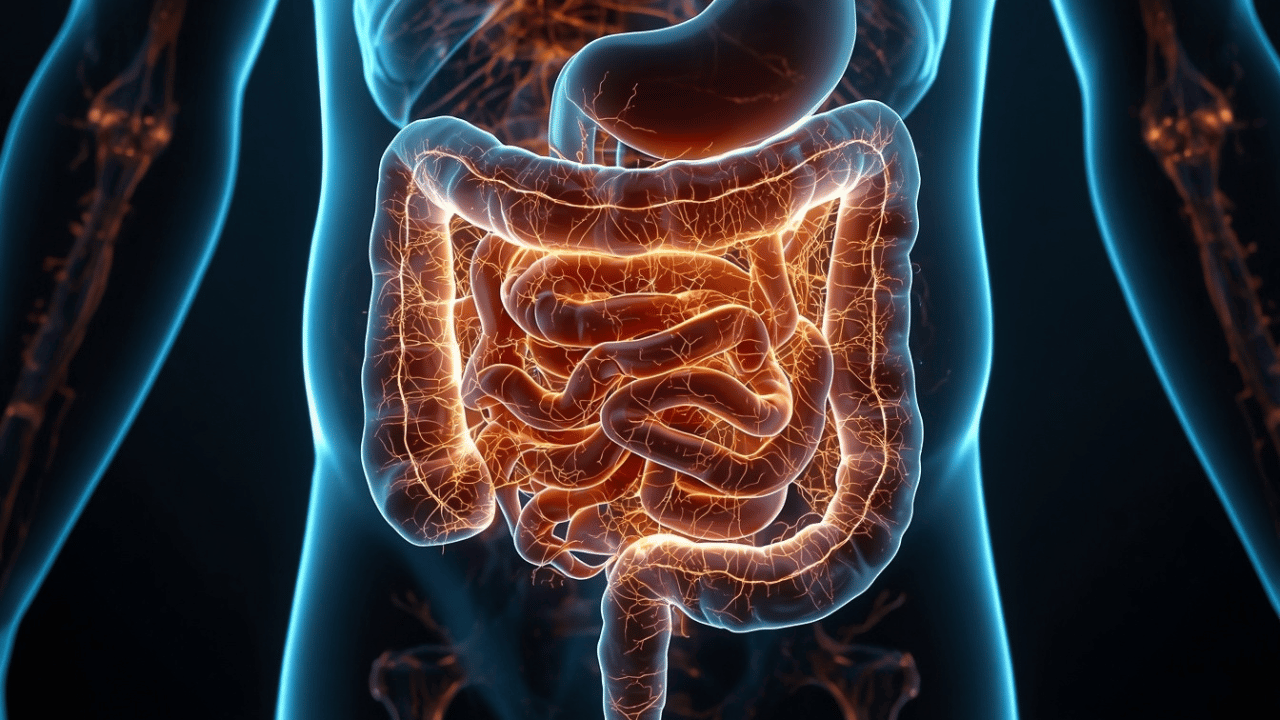 Detailed 3D visual mapping of the Enteric Nervous System embedded within the intestinal tissue layers, communicating with the central nervous system via the vagus nerve.
Despite its impressive independence, the Enteric Nervous System maintains a continuous, high-speed communication channel with the central nervous system. This connection is maintained primarily by the Vagus Nerve, the tenth cranial nerve, which runs directly from the brainstem down to the abdominal organs. For decades, traditional medicine assumed that the brain primarily used this nerve to send commands down to the digestive tract. However, modern neuro-gastroenterology has revealed a revolutionary biological fact: up to ninety percent of the nerve fibers within the vagus nerve carry information upward from the gut to the brain.
This direct neural pipeline means that the operational health of your digestive system continuously dictates your central neurological state. When the gastrointestinal wall experiences localized cellular stress, inflammation, or microbial imbalance, the Enteric Nervous System detects these threat signals and sends high-volume warnings up the vagus nerve straight into the brainstem and limbic system. This continuous neuro-inflammatory signaling is a primary driver behind unexplained mood disturbances, chronic anxiety, systemic fatigue, and persistent brain fog.
6. The Complete Digestive Pathway: From Ingestion to Systemic Integration
To truly appreciate the elegance of human digestion, we must step back and observe how all these distinct anatomical components operate together as an unbroken, continuous system. Digestion is not a series of isolated events, but a synchronized physiological cascade where every single phase directly triggers and relies upon the success of the phase before it.
The journey begins in the head with the cephalic phase. Simply seeing, smelling, or thinking about food triggers signals from the central nervous system down through the vagus nerve, instructing the salivary glands to produce saliva and prompting parietal cells in the stomach to begin secreting hydrochloric acid before food even touches your lips. Once mastication and swallowing occur, the mechanical pressure of the bolus entering the stomach initiates the gastric phase, driving intense muscular churning and heavy acid release.
As the stomach gradually empties its contents into the duodenum, the intestinal phase takes command. The sudden arrival of acidic, lipid-rich chyme triggers immediate hormonal cascades. Enteroendocrine cells in the intestinal wall release secretin and cholecystokinin into the bloodstream. These chemical messengers simultaneously signal the pancreas to release neutralizing bicarbonate and digestive enzymes, instruct the gallbladder to contract and discharge concentrated bile, and tell the stomach to slow its emptying rate to allow adequate processing time.
Throughout the length of the small intestine, complex macronutrients are dismantled into their fundamental chemical building blocks. Proteins become amino acids, complex carbohydrates become simple monosaccharides, and fats become free fatty acids and monoglycerides. These basic molecules are absorbed across the microscopic brush border, entering either the blood capillaries or the lymphatic lacteals. The absorbed nutrients entering the bloodstream are routed immediately through the hepatic portal system to the liver, where toxic substances are filtered, glucose is converted into stored glycogen, and essential amino acids are redistributed throughout the body.
Finally, the remaining indigestible residues pass into the large intestine, where the native microbiome performs its vital metabolic fermentation. Water and essential electrolytes are carefully recovered into systemic blood circulation, while short-chain fatty acids nourish the colonic lining and reinforce systemic immune tolerance. The remaining consolidated waste is moved into the rectum, where stretch receptors trigger the defecation reflex, completing the extraordinary biological cycle of human digestion.
Conclusion: The Digestive System as a Systemic Health Anchor
The human digestive system is far more than a simple food processor or a mechanical waste canal. As clinical physiology and modern research continuously demonstrate, it functions as a highly integrated, self-regulating ecosystem that directly governs cellular energy, immune tolerance, hormone production, and neurological health.
From the biochemical precision of stomach acid and pancreatic enzyme delivery, to the massive absorptive surface of the small intestine and the complex metabolic output of the gut microbiome, every phase of digestion is linked in an uninterrupted chain. When one segment experiences dysfunction—whether through mucosal barrier degradation, enzymatic insufficiency, or impaired vagal signaling—the downstream effects resonate across the entire body, manifesting as chronic fatigue, metabolic imbalances, or systemic neuro-inflammation.
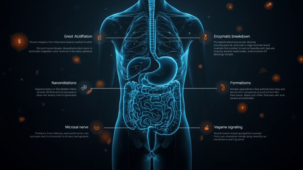 Comprehensive clinical infographic vector depicting the 5 key pillars of human digestive health, from gastric acid balance to gut-brain vagal signaling.
Ultimately, supporting digestive health requires looking beyond isolated symptoms and recognizing the gastrointestinal tract as the primary engine of human vitality. By prioritizing optimal nutrient breakdown, protecting intestinal barrier integrity, and nourishing the living gut ecosystem, we safeguard not just our digestion, but our entire systemic health.
💡 Key Takeaways: The Digestive Continuum at a Glance
- Continuous Biochemical Cascade: Digestion relies on a strictly ordered sequence where each phase (cephalic, gastric, intestinal) triggers the next. A failure at the top of the chain impacts all downstream absorption.
- Accessory Synergy: The liver, gallbladder, and pancreas provide the heavy chemical machinery—bile and pancreatic enzymes—essential for breaking down fats, proteins, and carbohydrates into absorbable units.
- Massive Absorptive Surface: Through plicae circulares, villi, and microvilli, the small intestine expands its surface area to roughly 200 square meters to maximize nutrient transport.
- The Microbiome Engine: Over 100 trillion microbes in the colon process indigestible fibers into short-chain fatty acids (like butyrate), which repair the colonic lining and suppress systemic inflammation.
- The Gut-Brain Axis: Containing over 500 million neurons, the Enteric Nervous System operates autonomously while transmitting up to 90% of its vagal nerve signals upward to the central nervous system, directly impacting mood, focus, and brain health.
Frequently Asked Questions (FAQ)
1. What are the main components of the human digestive system?
The human digestive system consists of two primary divisions: the Gastrointestinal (GI) Tract, which includes the mouth, pharynx, esophagus, stomach, small intestine, and large intestine; and the Accessory Organs, which include the teeth, tongue, salivary glands, liver, gallbladder, and pancreas.
2. What is the difference between mechanical and chemical digestion?
Mechanical digestion involves the physical breakdown of food into smaller pieces without changing its chemical structure, such as chewing in the mouth and churning in the stomach. Chemical digestion uses enzymes, acids, and bile to break complex food molecules down into simple, absorbable chemical nutrients.
3. How does the stomach protect itself from its own acid?
The stomach protects its muscular walls from highly acidic gastric juices using a specialized mucosal barrier. Gastric epithelial cells continuously secrete a thick layer of bicarbonate-rich mucus that neutralizes hydrochloric acid right at the cellular surface, preventing tissue self-digestion.
4. What is the primary function of the small intestine?
The small intestine is the primary site for both the final chemical breakdown of food and the absorption of nutrients into the bloodstream. Its specialized internal structures—plicae circulares, villi, and microvilli—maximize surface area to efficiently absorb amino acids, simple sugars, fatty acids, vitamins, and minerals.
5. Why are the liver, gallbladder, and pancreas called accessory organs?
They are classified as accessory organs because food does not physically pass through them. Instead, they produce, store, and secrete essential digestive chemicals, enzymes, and bile through specialized ducts directly into the small intestine to complete chemical digestion.
6. What role does bile play in digestion?
Bile, produced by the liver and stored in the gallbladder, contains specialized bile salts that act as detergents. Bile breaks large fat globules down into microscopic droplets through emulsification, vastly increasing the surface area for pancreatic lipase to break down dietary fats.
7. What is the Enteric Nervous System (ENS)?
The Enteric Nervous System is an independent neural network containing over 500 million neurons embedded directly within the tissue walls of the gastrointestinal tract. Often called the "second brain," it autonomously controls gut motility, fluid secretion, and localized blood flow.
8. How does the gut communicate with the brain?
The gut and brain communicate continuously through the gut-brain axis, primarily via the Vagus Nerve, circulating hormones, and microbial metabolites. Remarkably, up to 90% of the nerve fibers in the vagus nerve transmit sensory signals upward from the gut to the brain.
9. What happens in the large intestine?
The large intestine primary absorbs remaining water and essential electrolytes, converts liquid digestive waste into solid stool, and houses the gut microbiome, which ferments indigestible dietary fibers into beneficial short-chain fatty acids.
10. What is the gut microbiome and why is it important?
The gut microbiome is a vast ecosystem of trillions of microorganisms living primarily within the large intestine. It plays a critical role in fermenting fibers, producing vitamins, protecting against harmful pathogens, maintaining the intestinal mucosal barrier, and regulating systemic immune function.
11. What causes mucosal barrier breakdown or "leaky gut"?
Mucosal barrier breakdown occurs when the tight junction proteins connecting intestinal lining cells become damaged by chronic inflammation, poor dietary habits, stress, or toxin exposure. This allows unrefined food particles and bacterial endotoxins to leak into the bloodstream, triggering systemic immune responses.
12. How long does the complete digestive process take?
The entire digestive journey, from initial ingestion to final elimination, typically takes between 24 and 72 hours, depending on individual metabolic rate, hydration levels, physical activity, and dietary composition.
13. What is the function of the Migrating Motor Complex (MMC)?
The Migrating Motor Complex is a distinct electrical and muscular wave pattern that occurs in the stomach and small intestine during periods of fasting. It acts as an internal broom, sweeping undigested food debris and bacteria down into the colon to prevent small intestinal bacterial overgrowth (SIBO).
14. How does stress impact digestive physiology?
Psychological or physical stress activates the sympathetic nervous system ("fight or flight"), which diverts blood flow away from the gastrointestinal tract, slows down digestive motility, reduces enzyme and acid production, and can compromise the structural integrity of the intestinal lining.
15. How can I support optimal digestive function?
Supporting optimal digestion requires thoroughly chewing food, staying adequately hydrated, consuming a wide variety of fiber-rich whole foods to nourish the gut microbiome, managing chronic stress levels, and avoiding unnecessary overuse of medications that disrupt the gastric mucosal lining.
keep reading
Gut Health & Supplements
DigestiStart Review: Can This Herbal Formula Help Restore Digestive Health & Relieve Bloating?
Gut Health & Supplements
The Ultimate Guide to GERD: Causes, Symptoms & Natural Relief
Gut Health & Supplements